
1. Introduction: Why DGH A Matters
In modern ophthalmology, precise eye measurements are foundational for diagnosis, surgery planning, and monitoring conditions like myopia. Among the tools employed, DGH A has carved a niche for being portable, accurate, and user-friendly. This guide aims to unpack everything about DGH A — from its technical basis to real-world uses — so that doctors, students, and even curious lay readers can appreciate its value.
2. What Is DGH A? / Definition & Overview
- DGH A refers to a portable A-Scan ultrasound biometry device, notably the Scanmate A model by DGH Technology.
- It’s used to measure axial length, lens thickness, and other ocular dimensions via sound waves.
- DGH A belongs to the class of A-Scan ultrasound devices, which are standard in ophthalmic biometry.
- It is distinguished by being compact, lightweight, and designed for ease-of-use compared to older or larger machines.
So, “DGH A” is essentially a brand / model of eye measurement tool used primarily by eye care professionals.
Table of Contents
- Introduction: Why DGH A Matters
- What Is DGH A? / Definition & Overview
- History & Development of DGH A Technology
- How DGH A Works: Principles & Mechanism
- Key Components & Features of DGH A
- Uses & Applications of DGH A
- Benefits & Advantages of DGH A
- Limitations, Risks & Considerations
- DGH A in Comparison with Other Eye Measurement Tools
- Practical Guide: How to Use DGH A in Clinics
- Interpretation of Results & Clinical Significance
- Best Practices & Maintenance Tips
- Future Trends & Innovations in DGH A / Eye Biometry
- Case Studies & Real-World Examples
- Frequently Asked Questions (FAQs)
- Conclusion & Final Thoughts
3. History & Development of DGH A Technology
To appreciate DGH A, it’s useful to understand how biometry evolved:
- Early A-Scan ultrasound devices were large, bulky, and required extensive training.
- Over time, miniaturization, digital signal processing, and software improvements allowed more compact tools.
- DGH Technology (a company specializing in ophthalmic instrumentation) developed the DGH A / Scanmate A to bring precision into smaller, clinic- or portable-level devices.
- Regulatory approvals (FDA, CE) affirmed its safety and reliability in many markets.
Thus, DGH A stands on decades of ophthalmic measurement evolution, combining classic ultrasound principles with modern software.
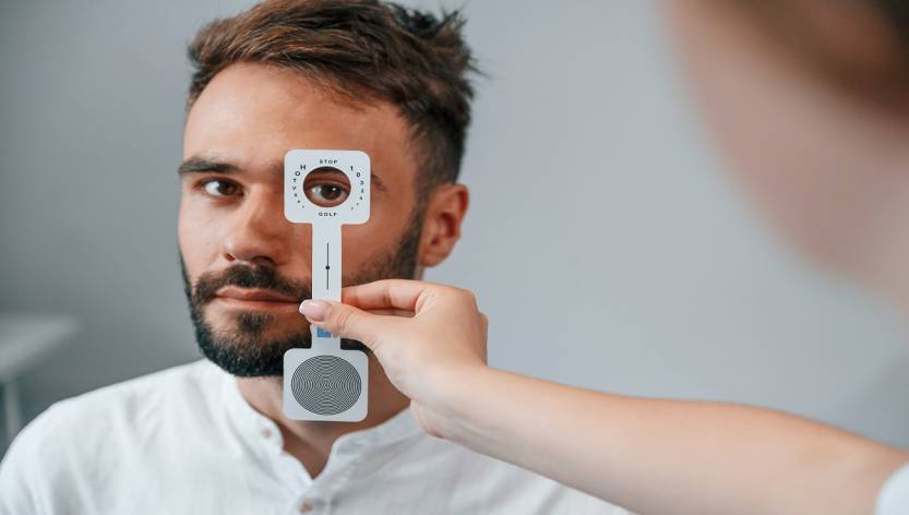
4. How DGH A Works: Principles & Mechanism
Understanding how DGH A measures the eye is crucial for interpreting its data correctly.
Basic Principle: Ultrasound A-Scan Biometry
- The “A-Scan” sends a short ultrasonic pulse along the optical axis of the eye.
- The pulse travels through ocular media (cornea, aqueous humor, lens, vitreous) and reflects (“echoes”) at boundaries with different acoustic impedances.
- The device measures the time delay between emission and return of echoes.
- Using known sound speed (in each medium), the system calculates distances (e.g. from cornea to retina = axial length).
Specifics in DGH A
- The probe typically uses ~10 MHz frequency (higher frequencies yield better resolution but shallower penetration).
- There may be contact mode (probe touches cornea) or immersion mode (probe submerged in fluid via a shell).
- DGH A incorporates guidance features — e.g., visual “stars” or auditory feedback — to help the operator position the probe correctly.
- It uses built-in formulas (e.g. SRK/T, Holladay, Haigis) to translate biometry into intraocular lens (IOL) power estimates in cataract surgery planning.
Safety & Calibration
- It uses harmless ultrasonic energy (non-ionizing).
- Many devices include safety locks (e.g. compression detection) to prevent damaging pressure on the eye.
- Regular calibration is needed to maintain measurement accuracy.
5. Key Components & Features of DGH A
Here are the core parts and notable features that make DGH A useful:
- Probe / Transducer — the handpiece that emits and receives ultrasound waves.
- USB Interface — connects to a computer to run the software.
- Software & User Interface — screen displays stars, graphs, measurement readouts, IOL formulas.
- Guidance System — visual (stars, cross-hairs) plus sound feedback to align the probe correctly.
- Safety Lock / Compression Lockout — prevents excessive force.
- Multiple Measurement Modes — contact vs immersion.
- IOL Calculation Engine — built-in formulas to compute lens power.
- Data Export / Reporting — ability to save, export, or print measurement results.
- Portability — compact size, lightweight construction (device under 1–2 lb).
- Certification & Compliance — FDA, CE marks for safety in many countries.
These features distinguish DGH A from more primitive A-Scans or large hospital machines.
6. Uses & Applications of DGH A
Where and how is DGH A used in practice?
- Cataract Surgery Planning
- Determining axial length and expected lens power (IOL) is critical for good postoperative refractive outcomes.
- Myopia / Eye Growth Tracking
- Measuring changes in axial length over time in children to monitor progression or therapy (e.g. orthokeratology)
- Ophthalmic Clinics / Hospitals
- Day-to-day biometry needs where space or budget is constrained.
- Mobile Eye Camps / Outreach
- Because it’s portable, it can be carried to rural or underserved locations.
- Post-LASIK / Post-Refractive Surgery Eyes
- For eyes that have changed shape, specialized formulas in DGH A help estimate lens power correctly.
- Research / Clinical Studies
- In ophthalmic research to gather biometric data across populations.
- Teaching / Training Purposes
- Because of ease of use, it may be used as a training tool for residents or students.
7. Benefits & Advantages of DGH A
Why might a clinic or ophthalmologist choose DGH A over alternatives?
- Portability & Compact Size
- Easy to move, deploy in remote settings, or aboard mobile clinics.
- User-Friendly Interface & Guidance
- Visual and auditory feedback reduce operator error and training time.
- Dual Mode (Contact & Immersion)
- Flexibility depending on patient condition or preference.
- Built-In IOL Formulas & Power Calculation
- Saves time and reduces manual calculation error.
- Cost-Effective for Smaller Clinics
- Less expensive than large full-featured biometry systems.
- Accuracy & Consistency
- Good repeatability if used properly.
- Safety Features
- Compression lock, handling safeguards.
- Regulatory Approvals
- Gives confidence in using it clinically (FDA, CE).
These advantages make DGH A a favorable compromise between performance and practicality.
8. Limitations, Risks & Considerations
No tool is perfect. Here are some caveats of DGH A:
- Contact mode error
- Touching the cornea may slightly compress or distort the eye, introducing small measurement errors.
- Operator dependence
- Proper technique (probe alignment, steadiness) matters greatly for accuracy.
- Not fully automated
- Some modern devices (e.g. optical biometers) do measurement more automatically / non-contact.
- Limitation in dense media
- In eyes with media opacities (e.g. dense cataract or corneal scars), ultrasound may struggle.
- Requires calibration & maintenance
- Over time, transducers may drift; quality assurance is essential.
- Limited by formula assumptions
- IOL formulas rely on assumptions; for highly abnormal eyes (extreme myopia, prior surgery), errors may increase.
- Comfort / Patient factors
- Contact mode requires patient cooperation; immersion mode needs shell and fluid, which some patients may find uncomfortable.
- Lack of other metrics
- Unlike more advanced devices, DGH A may not provide metrics like anterior chamber depth, corneal curvature, etc., all in one.
One should weigh these when selecting DGH A versus more advanced or integrated systems.
9. DGH A in Comparison with Other Eye Measurement Tools
Let’s compare DGH A with other common biometry tools:
| Device / Method | Strengths | Weaknesses / Compared to DGH A |
|---|---|---|
| Optical Biometer (e.g. IOLMaster, Lenstar) | Non-contact, high precision, more metrics (e.g. keratometry) | More expensive; can fail in dense cataracts; less portable |
| Conventional A-Scan (older models) | Proven, reliable | Larger, less user-friendly, slower |
| Scheimpflug / OCT-based devices | More structural imaging, depth information | Not always designed for pure axial length; may not replace ultrasonics in dense media |
| Immersion-only ultrasound | Very accurate, reduces corneal compression error | Bulky, needs dedicated shells and fluid, slower setup |
In many cases, DGH A offers a sweet spot: better portability and ease-of-use than older A-Scans, and more flexibility (able to work in dense media) than pure optical biometers.
10. Practical Guide: How to Use DGH A in Clinics
Here’s a step-by-step outline of typical usage and practical tips:
Setup & Preparation
- Connect the device via USB to a compatible Windows computer.
- Install / launch software, ensure drivers and firmware are updated.
- Enter patient info: patient name, eye (OD/OS), age, etc.
- Select measurement mode (contact or immersion).
Preparing the Eye
- For contact mode: instill topical anesthetic drops (if required), ensure cornea is moist, ask patient to fixate straight ahead.
- For immersion mode: place a suitably sized shell (e.g. a Prager shell) filled with sterile saline or coupling fluid around the eye, ensuring alignment.
Probe Handling & Measurement
- Hold the probe steadily, aligned with the visual axis.
- Move slowly until you see alignment cues (stars, crosshairs, or sound feedback).
- Avoid pressing too hard; let the safety lock stop excessive compression.
- Capture multiple readings (e.g. 3–5) and average to reduce error.
- Check consistency: if one reading deviates significantly, discard.
Reviewing & Saving Results
- The software shows axial length, lens thickness, and IOL power suggestions.
- Examine signal waveforms — good peaks should be well defined.
- Export or print results to patient file or EMR.
- Back up data regularly.
Quality Control & Calibration
- Run periodic checks with phantom / calibration tools if available.
- Monitor drift or inconsistencies over time.
- Update software / firmware when manufacturer releases patches.
- Clean probe, cables, and connectors carefully.
Tips & Best Practices
- Always ensure probe and coupling medium are clean.
- Use consistent technique across patients to minimize operator variation.
- When in doubt, repeat measurements.
- Maintain proper lighting, patient comfort, and stability.
- Train operators thoroughly.
These operational standards help maximize measurement accuracy and reliability.
11. Interpretation of Results & Clinical Significance
What do the numbers produced by DGH A mean, and how are they used?
Axial Length (AL)
- Measured in millimeters, representing the distance from corneal vertex to retinal surface.
- A key determinant of refractive error: longer AL often correlates with myopia; shorter AL may correlate with hyperopia.
- In surgical planning (e.g. cataract), accurate AL is essential to selecting correct IOL power.
Lens Thickness
- The dimension of the natural crystalline lens (in mm).
- Important in calculating IOL power and determining effective lens position (ELP) models.
- May vary with accommodation, aging, and lens changes.
IOL Power Estimates
- Using AL, lens thickness, corneal curvature (if available externally), the device uses formulas (SRK/T, Holladay, Haigis, etc.) to compute suggested intraocular lens strength (in diopters).
- Clinicians may compare multiple formula outputs, especially in complex cases (post-refractive surgery, axial extremes).
Monitoring Change Over Time
- In longitudinal usage, repeated AL measurements help monitor axial elongation (especially in pediatric myopia).
- Deviations or aberrant growth may flag interventions.
Recognizing Errors & Artifacts
- Peaks that are irregular or too noisy suggest poor signal quality — those readings may be inaccurate.
- Discrepant multiple measurements (one outlier) should be ignored.
- Always cross-check with clinical context (e.g. biometric plausibility, patient history).
Understanding results is as critical as capturing them — the best data can only help if interpreted well.
12. Best Practices & Maintenance Tips
To keep DGH A performing reliably, adopt these practices:
- Daily cleaning: wipe probe surfaces, connectors, use proper cleaning agents compatible with ultrasound transducers.
- Storage: keep in dry, dust-free, temperature-controlled environment; protect from shocks.
- Firmware / software updates: install official updates to fix bugs or improve performance.
- Calibration / validation: use phantom standards or reference measurements periodically.
- Operator training: retrain staff regularly to reduce technique drift.
- Record keeping: maintain logs of maintenance, calibrations, anomalies.
- Backup of patient data: export or sync data regularly to avoid data loss.
- Check accessories: ensure cables, USB ports, shells, coupling fluid are all in good condition.
With proactive maintenance, the DGH A can serve reliably for many years.
13. Future Trends & Innovations in DGH A / Eye Biometry
What’s next? Some likely directions:
- Integration with imaging modalities (e.g. combining A-Scan with OCT, Scheimpflug) for more comprehensive metrics.
- AI / machine learning enhancements: better signal processing, noise reduction, automatic error detection.
- Wireless / portable upgrades: probe-to-tablet wireless connection, battery operation.
- Cloud-based data sharing and telemedicine: upload data to cloud for remote analysis.
- Miniaturization improvements: even smaller, lighter devices for field use.
- More advanced formulas: new IOL power formulas that adapt to extreme eyes or prior surgeries.
- Better real-time guidance: augmented reality (AR) overlay to visually guide probe placement.
These trends may influence future versions of DGH A or competing devices.
14. Case Studies & Real-World Examples
Here are hypothetical or documented scenarios where DGH A made a difference:
Case A: Rural Cataract Camp
In a remote region with limited infrastructure, clinicians carried DGH A to a mobile eye camp. They successfully measured axial lengths and recommended IOL powers on site, reducing the need for patients to travel to urban centers. The portability allowed high patient throughput.
Case B: Pediatric Myopia Monitoring
A children’s eye clinic used DGH A to monitor annual axial length growth in young myopic patients. The consistent measurements helped clinicians assess efficacy of treatments (e.g. atropine drops, orthokeratology) and adjust plans accordingly.
Case C: Post-LASIK Cataract Patient
A patient who had LASIK previously needed cataract surgery. Using standard formulas was risky. The DGH A’s software allowed use of post-refractive formulas, enabling better IOL power estimates tailored to the altered corneal structure.
Case D: Small Clinic Upgrade
A small private ophthalmic practice upgraded from an older A-Scan to DGH A. They found the learning curve short, accuracy improvement noticeable, and patient throughput increased because of faster scans.
These illustrate how DGH A can play roles in diverse settings.
15. Frequently Asked Questions (FAQs)
Q1. What does “DGH A” stand for?
A: It’s a product name/model from DGH Technology (Scanmate A). The “A” denotes the A-Scan modality.
Q2. Is DGH A suitable for children?
A: Yes. It covers a broad measurement range (e.g. 15–40 mm), making it suitable for both pediatric and adult eyes.
Q3. Can DGH A replace optical biometers?
A: Not entirely. Optical biometers excel in non-dense media and can measure additional parameters. But in dense cataracts, DGH A (ultrasound) may succeed where optics fail.
Q4. How accurate is DGH A?
A: When used properly, it can deliver high repeatability (±0.03 mm or similar). But real accuracy depends on operator technique, calibration, and signal quality.
Q5. Does DGH A require special training?
A: Some training is essential, especially for probe handling, alignment, and interpreting waveforms. But its intuitive guidance features reduce the learning burden.
Q6. Can DGH A be used in patients after LASIK / PRK?
A: Yes. The device includes specific IOL formulas for post-refractive surgery eyes.
Q7. How often should the device be calibrated?
A: It depends on usage, but periodic calibration or validation (e.g. monthly or quarterly) is recommended, along with routine QC checks.
Q8. Is DGH A safe?
A: Yes. It uses non-ionizing ultrasound and has safety features (e.g. compression lock). When used properly, risks are minimal.
Q9. What are the constraints in dense cataracts?
A: While ultrasound can often penetrate better than optical methods, extremely dense or opaque media may still degrade signal quality or prevent accurate echo reflection.
Q10. How does ambient environment affect readings?
A: Vibration, movement, poor patient fixation, or improper coupling fluids can degrade measurement quality. Controlled environment and technique matter.
16. Conclusion & Final Thoughts
The DGH A (Scanmate A) offers a compelling option in ophthalmic biometry: portable, relatively affordable, and capable of clinically meaningful measurements. It bridges the gap between large, fixed devices and more rudimentary units.
That said, its value hinges on correct usage, quality control, and understanding its limitations. No device is perfect—optical biometers and multimodal devices will continue to push the frontier. But in environments where cost, portability, or media opacities pose challenges, DGH A shines.
Read More: hcooch ch2 h2o: Structure, Mechanism, and Key Applications