
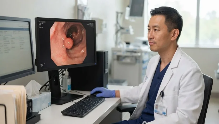
A tubular adenoma is not colon cancer. If you see this term on a colonoscopy report or patient portal, it means doctors found a type of polyp in the colon or rectum. They usually remove the polyp during the colonoscopy, and a lab examines the tissue under a microscope to identify its type.
That name scares people, and the wording on the report does not help. Adenoma and adenocarcinoma sit a few letters apart. One is a polyp. The other is cancer. They are not the same diagnosis.
This is general information, not a reading of anyone’s personal report. A doctor confirms a tubular adenoma after colonoscopy and pathology. Symptoms cannot.
What a tubular adenoma actually is
The colon lining is full of glands. Sometimes a cluster of those gland cells starts growing in a slightly disorderly way and forms a small bump that sticks into the bowel. That bump is a polyp. If the tissue looks like a bundle of tiny tubes under the microscope, the pathologist calls it a tubular adenoma. Doctors also say adenomatous polyp or colon adenoma.
The National Cancer Institute describes colorectal adenomas as common polyps made of gland-like tissue. They are not cancer. They are more likely to become cancer than some other polyps if they sit there for years, which is why they get removed.
Pathologists split conventional adenomas by architecture:
A tubular pattern is the usual one, especially in smaller polyps. Tube-shaped glands make up most of the growth, often more than about 75 percent. Among this family of polyps, tubular adenomas tend to carry the lowest chance of turning into cancer.
A villous pattern looks more like finger-like fronds. These are less common. The American Cancer Society notes that villous adenomas are more likely to change into cancer than tubular ones.
A tubulovillous adenoma is a mix.
People also see sessile or pedunculated on reports. Sessile means the polyp sits on a broad base. Pedunculated means it has a stalk. Those words describe the shape the endoscopist saw, not whether the tissue is malignant.
Colon polyps in general are ordinary findings in adults. NIDDK estimates that somewhere between 15% and 40% of adults have them. Mayo Clinic says about 1 in 3 people over 50 will develop at least one adenoma at some point. Most of those growths never make a person feel sick. A lot of them never would have become cancer. Screening finds them anyway, and that is largely the point.
Is it cancer? Is it “precancerous”?
It is not cancer.
It is called precancerous because some adenomas can slowly travel a recognized path toward colorectal adenocarcinoma. Textbooks call that the adenoma-carcinoma sequence: lining cells pick up genetic changes, a polyp forms, and a fraction of those polyps, left alone long enough, become cancer. The clock is usually measured in many years, not weeks.
Two things can be true at once, and both matter.
Most tubular adenomas never become cancer, particularly the small ones that come out whole. At the same time, most colorectal cancers start as polyps. Take the polyps out, and a large share of that risk leaves with them. That is why colonoscopy prevents cancer instead of only detecting it.
“Precancerous” describes a finding that may increase cancer risk. It is not a stage of cancer, and it does not mean a hidden tumor exists elsewhere. It also does not mean chemotherapy will be needed.
A few neighboring labels get mixed together online:
Hyperplastic polyps are common, especially in the rectum and sigmoid. Small ones there are usually not treated as precancers.
Sessile serrated lesions (also called sessile serrated polyps) are a different biology. They can also lead to cancer, just not by the classic tubular-adenoma route. They are not tubular adenomas.
High-grade dysplasia is still not invasive cancer. The cells look more abnormal, but they have not grown down into deeper tissue. Invasion is the line pathologists use for adenocarcinoma.
Colorectal adenocarcinoma is cancer. If that is on the report, the conversation is different from the one this article is about.
If the report only says tubular adenoma, the lab did not diagnose cancer in that polyp.
Why these polyps form
Nobody can point to one cause in a single person. NIDDK is blunt about it: experts are not sure what causes colon polyps. What doctors have are patterns.
Cells in the lining grow a little too fast and a little too sloppily. Over a long stretch of time, that extra tissue becomes a polyp. Age does a lot of the work. So do inherited risk, inflammation, and some of the same habits linked to colorectal cancer.
People are more likely to form polyps or to develop colorectal cancer if they are older, male, or already had adenomas in the past. A parent, sibling, or child with colorectal cancer or adenomatous polyps raises the odds, more so if that relative was diagnosed before 50 or if more than one close relative was affected. Long-standing ulcerative colitis or Crohn’s disease involving the colon raises risk too.
A handful of inherited conditions change the picture completely: Lynch syndrome, familial adenomatous polyposis (FAP), MUTYH-associated polyposis. They are uncommon. They can mean many polyps or a high lifetime cancer risk, and they do not follow the average-risk follow-up schedule.
Lifestyle associations include extra body weight, type 2 diabetes, smoking, heavier drinking, a lot of red or processed meat, and little physical activity. Associations are not destiny. Plenty of people with no dramatic risk factors still grow a small tubular adenoma. Plenty of people with risk factors never do. The list mainly helps decide when to start screening and how closely to watch afterward.
Most of them cause no symptoms
This is the part screening exists for.
NIDDK says most people with colon polyps feel well. You cannot tell they are there. Mayo Clinic’s tubular adenoma pages say the same: they grow slowly and tend to show up on a routine exam, not because something hurt.
When a polyp does cause trouble, it is usually because it is larger or because it has been bleeding. That can look like blood on the paper, red streaks in the stool, or stool that appears black. Some people become tired from iron-deficiency anemia after slow, unnoticed blood loss. A new change in bowel habits or belly discomfort can show up too.
Those symptoms have a long list of other explanations: hemorrhoids, a small tear, infection, inflammatory bowel disease. They do not prove an adenoma is present. They also do not prove one is absent.
New rectal bleeding or blood in the stool is a reason to call a doctor rather than wait for the next screening date. So is a change in bowel habits that does not settle, pain that hangs around, or weight loss nobody was trying for. Feeling fine is not a reason to skip screening. The polyps this test is best at catching are often silent.
How the finding usually happens
The gastroenterologist may already have mentioned a polyp on the day of the colonoscopy, once the sedation had worn off enough for a conversation. That is not the final diagnosis. The polyp goes to pathology. Results often take about a week or two, depending on the lab. Some people get a portal result first and a phone call later. The wait is ordinary. It is also the stretch when most of the late-night searching happens.
In the U.S., average-risk screening now starts at 45. The U.S. Preventive Services Task Force, the American Cancer Society, and CDC all use that age. Screening often continues through 75; after that the decision gets more individual. People with inflammatory bowel disease, a personal or family history of colorectal cancer or certain polyps, or a known inherited syndrome may need to start earlier or come back more often.
Colonoscopy is the test that can find a polyp and remove it in the same session. Stool tests (FIT each year, or a stool DNA–FIT test about every three years), sigmoidoscopy, and CT colonography can also be used for screening. An abnormal result on those tests is not a polyp diagnosis. It is a reason to do a colonoscopy.
During colonoscopy, the doctor advances a camera through the whole colon. Quality matters more than patients are told in the waiting room. Did the scope reach the cecum? Was the bowel clean enough to see lesions bigger than 5 millimeters? Those details feed the later decision about when to come back.
If a polyp is there, it can often be lifted off with a wire snare or forceps. That is a polypectomy. Most people do not feel it. The piece of tissue is what the pathologist reads. The name “tubular adenoma” is a microscope name, not a camera name.
Reading the pathology report without panicking
Reports are written for the next clinician, not for a 2 a.m. Google session. A few lines do most of the work.
Tubular adenoma means adenomatous polyp with a mostly tubular pattern. Benign. Precancerous. Not cancer.
Size usually appears in millimeters. Ten millimeters is about a centimeter. That number is a common cutoff in follow-up guidelines. A 3-millimeter polyp and a 14-millimeter polyp can share a name and still be handled differently afterward.
Location may say cecum, ascending, transverse, descending, sigmoid, or rectum. Useful for the doctor. Not a cancer grade.
Dysplasia is the word that sends people to forums. It means the cells look abnormal but have not invaded. Conventional adenomas show dysplasia by definition, so seeing it on the report is expected, not a surprise twist.
Low-grade dysplasia is the milder, far more common version in small tubular adenomas. If that polyp came out completely, the risk from that growth is very low.
High-grade dysplasia is a more advanced precancerous change. Still not invasive cancer. Complete removal matters a great deal here, and the next colonoscopy is usually sooner. The American Cancer Society says high-grade dysplasia may move the follow-up date up; it is not, by itself, a cancer diagnosis.
Margins show up when the polyp came out in one piece. Negative margins means the edges looked clear. Positive or “cannot be determined” can mean a closer look, sometimes an earlier repeat exam. Tiny polyps removed with forceps may not get a dramatic margin discussion at all.
The ACS line that is easy to miss: the most important practical questions are whether the polyp was removed completely and whether it shows cancer. The tubular-versus-villous detail still matters, mainly because it helps set the next interval.
If a sentence on the report is opaque, the person to interpret it is the doctor who did the colonoscopy. They know whether the prep was poor, whether a piece was left, whether five other polyps were also taken out. The PDF in the portal does not.
What actually changes the cancer risk
Risk is not a single percentage attached to the phrase “tubular adenoma.” Online articles love a number. Real reports are a pile of features.
Size is one. Larger adenomas are more likely to hold advanced changes. In U.S. surveillance guidance, 10 mm is the line that usually pushes an adenoma into a higher-risk group even if it is still tubular.
Number is another. One or two small tubular adenomas is a different situation from a colon that grew several of them by the same exam.
Architecture matters. Tubular is lower risk than villous or tubulovillous.
Dysplasia grade matters. High-grade is an advanced feature.
And completeness of removal matters as much as any of those. A small tubular adenoma with low-grade dysplasia, taken out whole, is not the same clinical object as a large polyp removed in fragments with an uncertain edge.
Gastroenterologists use advanced adenoma as a shorthand for an adenoma that is 10 mm or larger, has villous or tubulovillous features, or shows high-grade dysplasia. That definition appears in the 2020 U.S. Multi-Society Task Force recommendations. Advanced is not a synonym for malignant. It is a reason to watch more closely.
Having formed one adenoma also means the colon is capable of forming others later. That is the actual argument for surveillance. People with one or two small, fully removed tubular adenomas generally have a lower chance of later advanced findings than people who already had an advanced adenoma or a handful of polyps.
Villous-adenoma statistics do not transfer onto a small tubular adenoma. If a search result is quoting cancer risk for a large villous growth, it is talking about a different polyp.
Treatment is usually the removal that already happened
There is no pill that melts these. There is no chemo protocol for a tubular adenoma without cancer.
The treatment is polypectomy. Small adenomas often come out in one piece. Larger ones may be taken off in several pieces, called piecemeal resection. Very large or awkwardly placed lesions sometimes need a more specialized endoscopic method, a second colonoscopy, or, uncommonly, surgery if the polyp cannot be removed safely through the scope.
If the endoscopist is not sure the entire growth is gone, the plan may include a short-interval repeat look at that spot. Leftover tissue can grow back. That is not “the cancer returning.” It is residual polyp.
Once pathology confirms a benign tubular adenoma and removal looks complete, the medical job shifts to the next exam date. It does not shift to oncology.
Afterward: the same polyp versus a new one
A polyp that was entirely removed does not grow back. People hear “they can come back” and assume the original growth regenerates like a weed. Two different things get lumped together.
If a bit of the polyp was left, that tissue can regrow at the same site. After piecemeal removal of a large lesion, U.S. guidance calls for an early check of the scar. For an adenoma 20 mm or larger taken out in pieces, the 2020 USMSTF recommendation is a colonoscopy in about 6 months to inspect that site.
Separately, a person who has grown adenomas can grow new ones later, somewhere else in the colon. Follow-up is for both problems.
There is no universal “next colonoscopy in X years” that fits every tubular adenoma. The interval depends on how many were found, how big they were, whether any had villous features or high-grade dysplasia, whether removal was complete, whether the bowel was clean, and whether the person has a family history, inflammatory bowel disease, or another reason to be in a higher-risk lane.
The 2020 USMSTF update is what many U.S. gastroenterologists use for average-risk adults after a high-quality exam. High-quality, in that document, means the scope reached the cecum and the prep was good enough to find lesions larger than 5 mm. Those recommendations are not a blanket rule for hereditary syndromes, inflammatory bowel disease, a malignant polyp, prior colorectal cancer, serrated polyposis, or a family history of colorectal cancer. In those settings the interval is often shorter.
For the most common report one or two tubular adenomas smaller than 10 mm, fully removed the U.S. range is often 7 to 10 years. That is longer than older advice, which used to land closer to 5 to 10 years. Three or four small tubular adenomas often land at 3 to 5 years. Five to ten small ones, any adenoma 10 mm or larger, villous/tubulovillous histology, or high-grade dysplasia: typically 3 years. More than ten adenomas at one exam: 1 year, and genetic evaluation may be discussed.
European groups sometimes send people with one or two small low-grade adenomas back to routine screening instead of keeping them on colonoscopy surveillance. Guidelines disagree. The interval that applies to a given person is the one their clinician chose after seeing the actual exam.
Staying at a healthy weight, moving more, limiting red and processed meat, not smoking, and keeping alcohol down may lower later risk. They do not replace the follow-up colonoscopy.
When to pick up the phone
Ask the doctor who did the procedure, or the clinician who ordered it, to translate the report if the size, number, dysplasia line, or margins are unclear. That is a normal request. So is asking why the next exam is in three years rather than ten, or the reverse.
Contact your doctor sooner if they could not remove the polyp completely, removed it in pieces, or were unsure whether they removed it all. Also speak with your doctor if you have a strong family history of colorectal cancer, several polyps, inflammatory bowel disease, or a known hereditary syndrome.
Do not ignore rectal bleeding, black stools, lasting changes in bowel habits, ongoing abdominal pain, or unexplained weight loss just because doctors removed your polyps. If you notice new bleeding, talk to your doctor instead of assuming hemorrhoids caused it.
In the U.S., major health groups recommend colorectal cancer screening for average-risk adults starting at age 45. People with a strong family history may need to start screening earlier, so they should discuss the right age with their doctor.
Common questions
Is a tubular adenoma dangerous?
The word is scarier than the usual finding. It is not cancer. A small, fully removed tubular adenoma with low-grade dysplasia is, for most people, a successfully prevented problem. Danger rises when a larger polyp, villous features, high-grade dysplasia, incomplete removal, or missed follow-up enter the picture.
Is it precancerous?
Yes. Adenomas may slowly develop into colorectal cancer if they remain in the colon. Precancerous is not the same as malignant.
Can it turn into cancer?
Some adenomas can slowly develop into cancer if doctors do not remove them. A small tubular adenoma with low-grade dysplasia is on the low end of that spectrum. Larger size, villous architecture, and high-grade dysplasia sit higher. Taking a benign adenoma out completely removes the risk from that polyp.
What size is concerning?
Doctors remove adenomas when they can do so safely. Ten millimeters (about 1 cm) is the guideline size that usually changes follow-up. A smaller tubular adenoma is still precancerous. It is typically lower risk if there were only one or two and they came out whole.
Do they come back?
If doctors remove the original polyp completely, it should not grow back. But if some tissue remains, it can grow again. New adenomas can also develop later, which is why follow-up exams are important.
How are they removed?
Usually during colonoscopy with a snare or forceps. Large polyps may need piecemeal removal or a more advanced endoscopic procedure. Surgery is the exception.
What happens after removal?
Pathology reads the tissue. If there is no cancer and removal looks complete, the next step is a surveillance interval, not cancer treatment.
Does this increase colorectal cancer risk?
It means the colon has already made a precancerous polyp, so later checks are in order. After removal, your risk depends on a few things: how many adenomas you had, their size, whether any had concerning features, whether doctors removed them all, and your personal or family history. One or two small tubular adenomas is generally a low-risk version of this story.
Does it mean colon cancer is coming?
No. Most people with a removed tubular adenoma do not go on to develop colorectal cancer, especially if they keep the follow-up their doctor actually recommended rather than the interval a search result guessed.
What if a stool test was positive?
Then colonoscopy is the next test. A positive stool test is not a diagnosis of adenoma or cancer.
If the report is sitting in a portal and the wording still feels off, take it to the clinician who has the colonoscopy note. They can say whether this was one 4-millimeter polyp or something that needs a closer calendar. That distinction is the whole clinical story. The phrase “tubular adenoma” is only the starting label.